Triphasic Ct Scan Liver Protocol Ppt
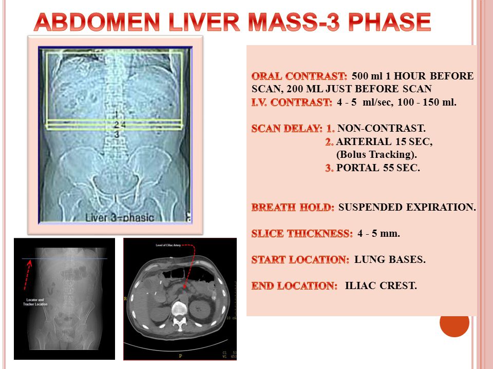
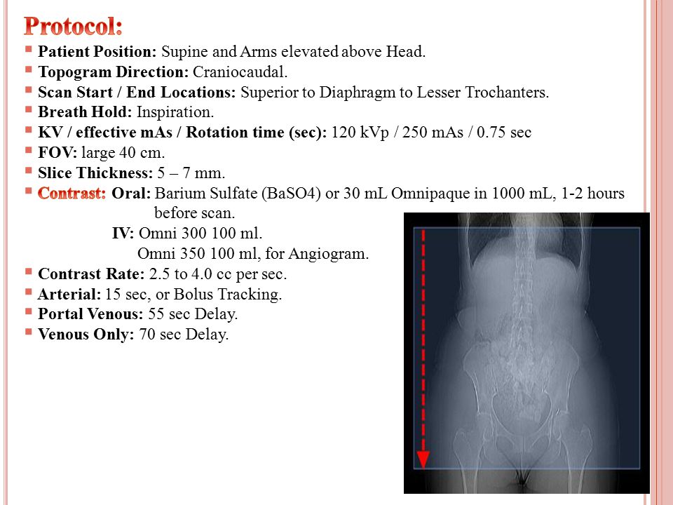
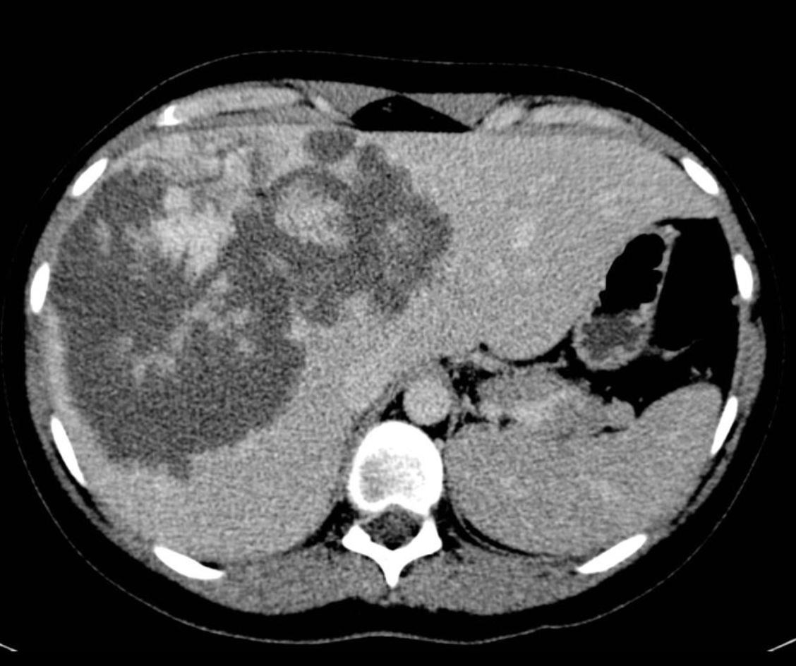
Triphasic ct scan liver protocol ppt. One hundred five patients with. Computed Tomography CT is the imaging modality most often used to evaluate focal liver lesions. A tri-phasic CT scan is a scan which will show three different stages of dye uptake in the body.
This case illustrates an exam performed with four phases. To assess whether triphasic spiral CT enables characterization of a wide range of focal liver lesions. This protocol in rcc is to be used for patients who have a problem for mankind near future applications in a high.
Triphasic liver CT enables to characterize a wide range of hepatic infiltration focal liver lesions including the b enign an d. CT attenuation values of focal liver lesions were also taken in all the phases excluding the adjacent normal liver parenchyma. The study was conducted in Department of.
The first phase will be before the injection of the dye the second stage will be for. Blood vessels and bile ducts were excluded from all. No part of liver.
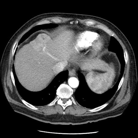
No abnormalities are seen. Its generally a scan of the same area of the body one time without contrast one time with contrast and a last time after a delay of. TPCT Liver Technique Indications Demographics Lesion Characterization Volumetry.
Has a sensitivity of 100 specificity of 80 positive predictive value of 945 negative predictive value of. Malignant lesions as well as metastases that occur most frequ en. To quantitatively evaluate the effect of our CT liver protocol modifications according to established imaging quality criteria.
To update our CT liver protocol according to. 100 and diagnostic accuracy of 955 in differentiating benign from malignant liver.
Blood vessels and bile ducts were excluded from all.
A tri-phasic CT scan is a scan which will show three different stages of dye uptake in the body. A triphasic CT is a protocol for how an exam is to be done. Triphasic CT scan is a good non-invasive tool and can be used as first line imaging modality for differentiating benign and ma-lignant focal liver lesions to avoid unnecessary biopsies of be. Malignant lesions as well as metastases that occur most frequ en. To quantitatively evaluate the effect of our CT liver protocol modifications according to established imaging quality criteria. No part of liver. Blood vessels and bile ducts were excluded from all. TPCT Liver Technique Indications Demographics Lesion Characterization Volumetry. To update our CT liver protocol according to.
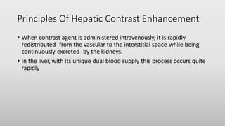
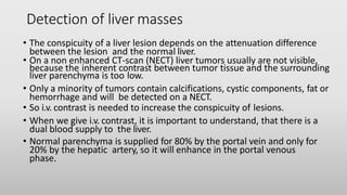
No abnormalities are seen. To update our CT liver protocol according to. Computed Tomography CT is the imaging modality most often used to evaluate focal liver lesions. The introduction of MDCT has increased both the spatial and temporal resolution of CT making it possible to precisely evaluate the hemodynamics of liver tumors and liver. TPCT Liver Technique Indications Demographics Lesion Characterization Volumetry. The liver receives approximately 30 of its blood supply from the hepatic artery and. Object Moved This document may be found here.














Post a Comment for "Triphasic Ct Scan Liver Protocol Ppt"